
Article by Vivek Radhakrishnan
Why do we need Testis Tissue Engineering?
Imagine you are consumed with cancer at a very young age termed as “Pediatric Cancer” and must undergo chemotherapy and radiation. There is a very high chance of the chemotherapeutics being gonadotoxic – toxic to your sex organs – leading to infertility. As per recent estimates, 80% of young adults suffering from pediatric cancer lead a cancer free life when treated with currently available cancer therapies and amongst them 42 – 66% are left infertile. This is a grave problem as the currently available methods to preserve fertility in adults such as sperm banking aren’t quite applicable to teen boys as they are unable to produce sperm before attaining puberty. The currently available methods for fertility preservation amongst boys is banking pre and peri- pubertal testicular tissues containing spermatogonial stem cells (SSCs) in a lab setting prior to gonadotoxic cancer treatments. The ideology is to utilize these tissues to produce sperm from the SSCs either in-vitro for use in assisted reproductive therapies – technique known as In Vitro Spermatogenesis (IVS) – or in-vivo upon auto transplantation. The below given figure explains the various use of these testicular tissues to aid in fertility. This technology has shown efficacy in generating sperm cells in rat upon simple air-medium interface culturing and in non-human primates upon subcutaneous implantation – houses the risk of reintroducing cancerous cells if present within the testis tissue – but there are no currently available models to grow the cryopreserved testis tissues into fully functional sperms in vitro.
This is where the field of tissue engineering steps in—with a interesting idea:
What if we could build a prepubertal human testis tissue model from human induced pluripotent stem cells (hIPSCs) to produce fully functional sperm cells using the patient’s own cells?
This ideology was conceived due to poor availability of human prepubertal patient tissues and to bridge the gap between animal and human models. Researchers are inclined towards using human induced pluripotent stem cells as they represent a robust source of human testis cells and can be used in patients whose testis tissue have low SSCs and cannot produce mature sperm due to genetic or pathological disorders. hIPSCs due to their early developmental state can be used to model human prepubertal tissue to facilitate technological development in the pursuit of human IVS.
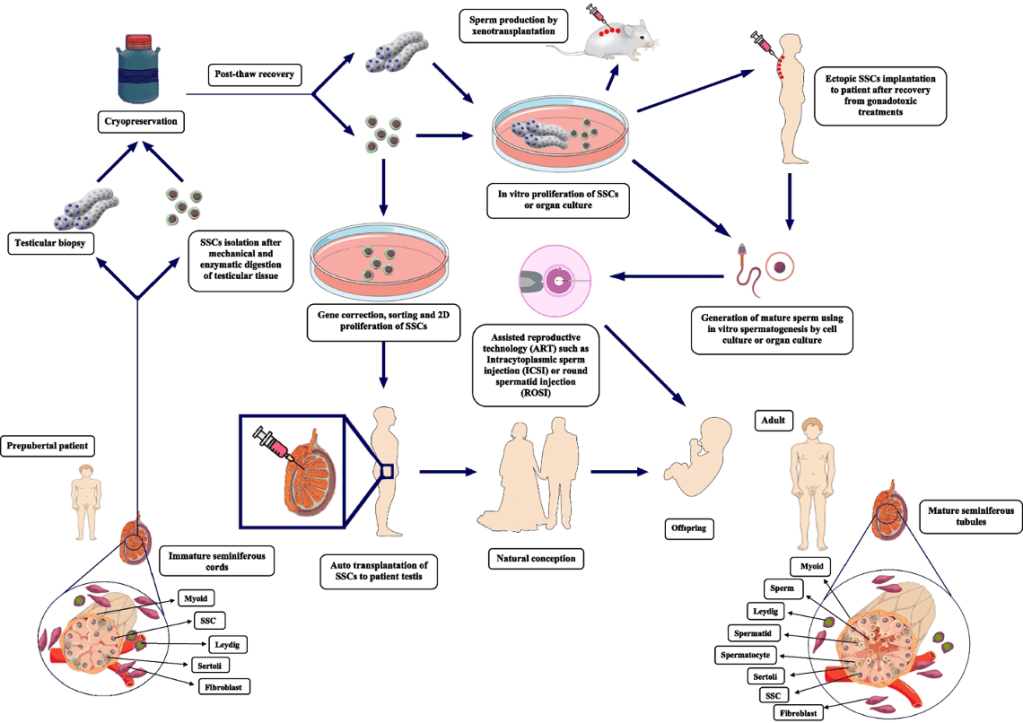
Figure 1: Currently available methods of utilizing prepubertal testis tissues for achieving fertilization in future
How do you build the Testis?
The study by MA Robinson et al., utilized an innovative 3D coaxial bioprinting to recapitulate the tubulerized, compartmentalized structure of native testis and create a more physiologically relevant model. The scaffold was generated using a coaxial printhead to generate a core-shell fiber system that represented the interstitial and tubular compartments of the testis cytoarchitecture. The prepubertal human testis was generated by seeding testicular cells differentiated from hIPSCs derived from red blood cells. The advantage of such a system is to genetically modify the testicular cells, this is of importance when generating models of patients who are pre-disposed to genetic disorders that induces infertility. This makes this model versatile and personalized. Along with generation of SSCs, Leydig cells, peritubular myoid cells and Sertoli cells were obtained from IPSCs upon a mesodermal differentiation. This was done to bring out cellular heterogeneity within the construct.
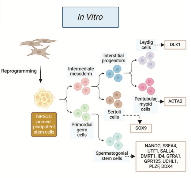
Figure 2: hIPSCs differentiation protocol
What did the research find ?
🧱 Structural Mimicry
3D Bioprinted Coaxial Core–Shell Constructs Recreate Native Architecture
- Seminiferous Cord Architecture: Using coaxial 3D bioprinting, Robinson et al. engineered a core–shell hydrogel structure that emulates the seminiferous tubules surrounded by interstitial space.
- Core: Encapsulated Sertoli cells and spermatogonial stem cell (SSC)-like cells
- Shell: Contained peritubular myoid-like cells and Leydig-like cells
- Bioink Composition:
- Core Ink (YIGSR peptide functionalized): Mimics laminin-rich basal lamina to promote Sertoli–SSC interaction and cord formation.
- Shell Ink (COL peptide functionalized): Mimics collagen matrix to enhance Leydig cell function.
- Dimensional Accuracy: Printed tubule diameters (~275 μm) and shell thickness (~500–600 μm) approximate human seminiferous cords and interstitial spacing.
- High Cell Viability: Bioprinting conducted under gentle conditions preserved cell viability and ensured reproducibility.
Comparison to Prior Models:
- Earlier organoid models and organ cultures lacked consistent structural arrangement.
- This coaxial model offers greater anatomical fidelity and reproducibility over self-organized organoids and rodent explant models.
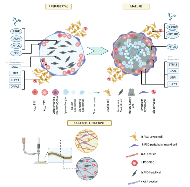
Figure 3: Key cell types and gene expression in prepubertal vs. mature testis (top), and the hiPSC-derived core–shell bioprinted model (bottom), using COL and YIGSR peptides to mimic native ECM and recreate the testis’s bicompartmental structure.
🧬 Biological Mimicry
hIPSC Differentiation into Testis Lineages
- Cell Source: Human induced pluripotent stem cells (hIPSCs) derived from adult somatic cells.
- Embryonic Lineage Simulation:
- Directed differentiation mimicked primordial germ cell (PGC) and intermediate mesoderm lineages.
- Yielded multiple testicular lineages via modulated signaling pathways:
- SSC-like cells: Expressed markers like NANOG, UTF1, PLZF—resembling pre-spermatogonia.
- Sertoli-like cells: Expressed SOX9, confirming their identity as nursing cells.
- Myoid-like cells: Expressed ACTA2 (α-SMA), indicating smooth muscle-like contractile identity.
- Leydig-like cells: Expressed DLK1, a known progenitor marker for testosterone-producing cells.
Single-cell RNA sequencing & immunostaining validated identity and purity of derived cell populations, matching native testicular cell marker profiles. This model thus recapitulated cellular diversity within tissue construct, a key milestone in tissue engineering.

Figure 4: Construct upon immunostaining for markers of maturation. It can be noted that cells are localized to either core or shell as per figure 3 and mature independently post 10 days of culture to give a mature diverse testis tissue.
⚙️ Functional Capability
In Vitro Puberty Simulation & Hormonal Responsiveness
- Sustained RA Delivery for Meiosis Induction:
- Used RA-loaded polycaprolactone microspheres for localized and prolonged retinoic acid delivery, mimicking in vivo stability and availability.
- Hormonal Milieu Emulation:
- Supplemented with FSH to stimulate Sertoli cell maturation.
- Likely used LH analog to activate Leydig steroidogenesis and testosterone production.
- Tissue Culture Outcomes:
- Early molecular and structural reorganization observed within 7 days in culture.
- Enabled study of early events in spermatogenesis onset under “pubertal-like” conditions.
What are the limitations and what is next ?
Despite its innovation, the 3D bioprinted testis model has yet to achieve full human spermatogenesis, as cultures lasted only 7–10 days and germ cells did not progress beyond early stages. Achieving complete meiosis and spermiogenesis will require extended culture times and potentially additional cues like cyclic retinoic acid pulses or physical stimulation. Sertoli cells showed partial maturation, but key features like gap junction formation were lacking, suggesting a need for prolonged hormone exposure or enhanced co-culture conditions. The absence of vascular and immune components limits nutrient delivery and mimicking of the native testis microenvironment; future versions may benefit from incorporating endothelial and testis-specific immune cells. Variability in hiPSC differentiation remains a challenge, requiring improved protocols to yield consistent germ and somatic cell populations across lines. Functionally, the next step is to drive germ cell progression into meiosis using refined culture conditions and signaling cues. Beyond fertility preservation, this model holds potential for studying human testis biology, toxicology, male contraception, and eventually generating sperm in vitro for assisted reproduction—pending rigorous validation for safety and genetic normalcy.
Conclusions
The 3D bioprinted coaxial testis model by Robinson et al. represents a significant advance in the quest for human in vitro spermatogenesis. By combining the developmental potential of hiPSCs with innovative bioprinting technology, the researchers created a structured, functional mimic of prepubertal testis tissue. This model demonstrated the importance of tissue architecture, cell–cell interactions, and sustained biochemical signaling (like retinoic acid delivery) in recapitulating early stages of sperm development and Sertoli cell maturation in vitro. While it has yet to produce human sperm, it lays crucial groundwork for future studies and brings us closer to a clinically viable solution for fertility restoration in young cancer survivors. This study outdid various existing functional models such as human testis on chip, organoid models and usage of rodent testis explants for achieving full spermatogenesis. Thus, this model helped bridge structural precision and endocrine responsiveness, thereby enabling a robust in vitro modeling of human testicular maturation. This model is a compelling proof of concept that holds promise in preserving fertility and in future could also be utilized as an alternative male fertility treatment as it relies on human induced pluripotent cells for spermatogenesis.
References:
- Salem, M., Khadivi, F., Javanbakht, P. et al. Advances of three-dimensional (3D) culture systems for in vitro spermatogenesis. Stem Cell Res Ther 14, 262 (2023). https://doi.org/10.1186/s13287-023-03466-6 *Introduction Image*
- Robinson, Meghan A., et al. “3D Bioprinted Coaxial Testis Model Using Human Induced Pluripotent Stem Cells: A Step Toward Bicompartmental Cytoarchitecture and Functionalization.” Advanced Healthcare Materials (2025): 2402606. *Article of discussion*
- Topraggaleh, T. R., Valojerdi, M. R., Montazeri, L., & Baharvand, H. (2019). A testis-derived macroporous 3D scaffold as a platform for the generation of mouse testicular organoids. Biomaterials science, 7(4), 1422-1436. *Cover Image*

Leave a comment